
Журнал «Медицина неотложных состояний» Том 21, №7, 2025
Вернуться к номеру
Оптимізація КТ-візуалізації при вогнепальних пораненнях: акцент на діагностичну точність
Авторы: E.M. Khoroshun (1, 2), V.V. Nehoduiko (1, 2), V.V. Makarov (1, 2), K.S. Smelyakov (3), A.S. Chupryna (3), Ye.V. Vakulik (3)
(1) - Military Medical Clinical Center of the Northern Region, Kharkiv, Ukraine
(2) - Kharkiv National Medical University, Kharkiv, Ukraine
(3) - Kharkiv National University of Radio Electronics, Kharkiv, Ukraine
Рубрики: Медицина неотложных состояний
Разделы: Клинические исследования
Версия для печати
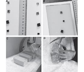
Актуальність. Удосконалення зображень, отриманих за допомогою комп’ютерної томографії (КТ), має особливе значення для ефективної діагностики вогнепальних поранень. Мета: розробити моделі, методи й тестове програмне забезпечення для градаційної корекції, що забезпечують автоматичне поліпшення рентгенівських та КТ-зображень у режимі реального часу, навіть при низькій видимості об’єктів малої щільності, як-от картон, шкіра та пластик. Матеріали та методи. Під час експериментів отримано КТ-зображення з неметалевими фрагментами різних типів, розташованими в окремих органах. Усі фрагменти були детально виміряні та задокументовані незалежними експертами для подальшого аналізу. Результати оцінювали шляхом чисельного та візуального порівняння з використанням традиційних і сучасних методів комп’ютерної обробки зображень. Результати. На основі отриманих даних створено уніфіковану модель, методику й тестове програмне забезпечення для градаційної корекції рентгенівських та КТ-зображень. Цей інструментарій дозволяє значно поліпшити якість зображень, підвищуючи їх контрастність до 5 разів порівняно з базовим зображенням при часі обробки одного зображення менше за секунду. Такі рішення є універсальними для різних форматів файлів, автоматично адаптуються до щільності матеріалів і забезпечують ефективну обробку даних. Висновки. Розроблені модель, методика й програмне забезпечення повністю відповідають поставленій меті — дають змогу швидко та якісно поліпшувати рентгенівські та КТ-зображення. Це дозволяє ефективно й точно діагностувати вогнепальні та осколкові поранення незалежно від типу сторонніх тіл у рані. Ефективність рішень підтверджена результатами експериментів.
Background. Improving computed tomography (CT) images for effective diagnosis of gunshot wounds is of particular importance. The aim of the work is to develop models, methods and test software for gradation correction that provides automatic enhancement of X-ray and CT images in real time, even under conditions of limited time and low visibility of low-density non-metallic objects, such as cardboard, leather and plastic. Materials and methods. During the experiments, CT images were obtained with non-metallic fragments of various types located in certain organs. All fragments were measured in detail and documented by independent experts for further analysis. The results were evaluated by numerical and visual comparison using traditional and modern computer image processing methods. Results. Based on the obtained data, a unified model, methodology and test software for gradation correction of X-ray and CT images were created. This toolkit allows to significantly improve the quality of images, increasing their contrast by up to 5 times, with a processing time for one image of less than 1 second. The developed solutions are universal for different file formats, automatically adapt to the density of materials and ensure efficient data processing. Conclusions. The developed model, methodology and software fully meet the set goal: they provide fast and high-quality improvement of X-ray and CT images. This allows for effective and accurate diagnosis of gunshot wounds, regardless of the type of foreign bodies in the wound. The effectiveness of the solutions is confirmed by the results of experiments.
комп’ютерна томографія; поліпшення зображень; градаційна корекція; діагностика вогнепальних поранень; ефективність
computed tomography; image enhancement; gradation correction; gunshot wound diagnosis; efficiency
Для ознакомления с полным содержанием статьи необходимо оформить подписку на журнал.
- Lurin I, Vorovskiy O, Makarov V, Khoroshun E, Nehoduiko V, et al. Management of thoracoabdominal gunshot injuries by using minimally invasive surgery at role 2 deployed field hospitals in Ukraine. BMC Surg. 2024;24(1). doi: 10.1186/s12893-024-02475-3.
- Panetta K, Kezebou L, Oludare V, Agaian S, Xia Z. TMO-Net: a parameter-free tone mapping operator using generative adversarial network and performance benchmarking on large scale HDR dataset. IEEE Access. 2021;9:39500-39517. doi: 10.1109/access.2021.3064295.
- Zhao L, Abdelhamed A, Brown MS. Learning tone curves for local image enhancement. IEEE Access. 2022;10:60099-60113. doi: 10.1109/access.2022.3178745.
- Kojima S, Suetake N. Single image enhancement using gradient-norm-based tone curve for images captured in wide-dynamic-range scenes. IEEE Access. 2024;12:1839-1851. doi: 10.1109/access.2023.3348121.
- Wang J, Qiao L, Lv H, Lv Z. Deep transfer learning-based multi-modal digital twins for enhancement and diagnostic analysis of brain MRI image. IEEE/ACM Trans Comput Biol Bioinform. 2023;20(4):2407-2419. doi: 10.1109/tcbb.2022.3168189.
- Thalhammer J, Schulthei M, Dorosti T, Lasser T, Pfeiffer F, et al. Improving automated hemorrhage detection at sparse-view CT via U-Net-based artifact reduction. Radiol Artif Intell. 2024;6(4). doi: 10.1148/ryai.230275.
- Merkow J, Lufkin R, Nguyen K, Soatto S, Tu Z, Vedaldi A. DeepRadiologyNet: radiologist-level pathology detection in CT head images. arXiv. 2017. doi: 10.48550/arxiv.1711.09313.
- Ries A, Dorosti T, Thalhammer J, Sasse D, Sauter A, et al. Improving image quality of sparse-view lung tumor CT images with U-Net. Eur Radiol Exp. 2024;8(1). doi: 10.1186/s41747-024-00450-4.
- Krk A, Yaar S, Durmaz MO. Prognostic factors in craniocerebral gunshot wounds: analysis of 30 patients from the neurosurgical viewpoint. Ulus Travma Acil Cerrahi Derg. 2020;26(6):859-864. doi: 10.14744/tjtes.2020.89947.
- Gascho D. Lodged bullets on computed tomography: three classification procedures for the virtual investigation of bullets or their fragments that cannot be recovered from the living patient. Med Sci Law. 2020;60(4):245-248. doi: 10.1177/0025802420962697.
- Junno JA, Kotiaho A, Oura P. Post-mortem computed tomo–graphy in forensic shooting distance estimation: a porcine cadaver study. BMC Res Notes. 2022;15(1):103. doi: 10.1186/s13104-022-05997-2.
- Radiant Viewer. Available from: https://www.radiantviewer.com/en/.
- Zeng Y, et al. A 2.5D deep learning-based method for drowning diagnosis using post-mortem computed tomography. IEEE J Biomed Health Inform. 2023;27(2):1026-1035. doi: 10.1109/jbhi.2022.3225416.
- Cao W, Wu R, Cao G, He Z. A comprehensive review of computer-aided diagnosis of pulmonary nodules based on computed tomography scans. IEEE Access. 2020;8:154007-154023. doi: 10.1109/access.2020.3018666.
- Li M, et al. Computer-aided diagnosis and staging of pancreatic cancer based on CT images. IEEE Access. 2020;8:141705-141718. doi: 10.1109/access.2020.3012967.
- Smelyakov K, Hvozdiev M, Chupryna A, Sandrkin D, Martovytskyi V. Comparative efficiency analysis of gradational correction models of highly lighted image. 2019 IEEE International Scientific-Practical Conference Problems of Infocommunications, Science and Techno–logy (PIC S&T). 703-708. doi: 10.1109/picst47496.2019.9061356.
- Smelyakov K, Pribylnov D, Martovytskyi V, Chupryna A. Investigation of network infrastructure control parameters for effective intellectual analysis. 14th International Conference on Advanced Trends in Radioelecrtronics, Telecommunications and Computer Engineering (TCSET). Lviv-Slavske; 2018. 983-986. doi: 10.1109/tcset.2018.8336359.
- Hansen TW, Ripa RS. Advances in imaging techniques for assessing myocardial microcirculation in people with diabetes. Diabetes Ther. 2025;16:785-797. doi: 10.1007/s13300-025-01710-1.