Резюме
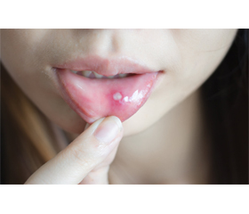
Актуальність. Узагальнюючої гіпотези стосовно етіології та патогенезу червоного плоского лишаю слизової оболонки порожнини рота на сьогодні не існує. Більшість авторів розглядають цей дерматоз як поліетіологічне захворювання, в етіології якого має значення метаболічна, нейроендокринна, вірусна природа, яка потенціюється токсикоалергічними, інфекційними та імунними тригерами. Мета дослідження: проаналізувати літературні джерела з характеристикою клітинного та видового складу мікрофлори слизової оболонки порожнини рота хворих на червоний плоский лишай. Матеріали та методи. Огляд та аналіз наукової й медичної літератури на основі баз даних Scopus, Web of Science, MedLine, PubMed, NCBI за останні 10 років, включно з оглядами літератури та результатами клінічних випробувань. Результати. Зміни у слизовій оболонці порожнини рота більшою мірою пов’язані із загальними патологічними процесами, що сприяють розвитку червоного плоского лишаю слизової оболонки порожнини рота. Таким чином, очевидною є необхідність раннього виявлення патологічних станів, які є факторами ризику розвитку червоного плоского лишаю слизової оболонки порожнини рота. Відсутність чітких уявлень про етіологію та патогенез вимагає всебічного обстеження із метою виділення найбільш ймовірного та вагомого провоканта у кожному окремому клінічному випадку. Висновки. Червоний плоский лишай є поліетіологічним захворюванням, що потрапляє в коло інтересів досить великої кількості фахівців, а саме імунологів, ендокринологів, неврологів, гастроентерологів, терапевтів та стоматологів. Оскільки дебют захворювання часто відбувається із ураження слизової оболонки порожнини рота, це вимагає від лікаря-стоматолога глибокого та системного клінічного мислення.
Background. There is currently no generalizing hypothesis regarding the etiology and pathogenesis of oral lichen planus. Most authors consider this dermatosis as a polyetiological disease of metabolic, neuroendocrine, viral nature, which is potentiated by toxic allergic, infectious and immune triggers. The purpose of the study is to analyze literary sources with the characteristics of the cellular and species composition of the microflora of the oral mucosa in patients with lichen planus. Materials and methods. Review and analysis of scientific and medical literature for the last 10 years was performed based on the Scopus, Web of Science, MedLine, PubMed, NCBI databases, including literature reviews and results of clinical trials. Results. Changes in the oral mucosa are mostly associated with general pathological processes that contribute to the development of oral lichen planus. Thus, the need for early detection of pathological conditions that are risk factors for the development of oral lichen planus is obvious. The lack of clear ideas about the etiology and pathogenesis requires a comprehensive examination in order to identify the most likely and significant trigger in each clinical case. Conclusions. Lichen planus is a polyetiological disease that generalizes the interests of quite a large number of specialists, namely immunologists, endocrinologists, neurologists, gastroenterologists, therapists and dentists. Since the onset of the disease often begins from damage to the oral mucosa, this requires deep and systematic clinical thinking from the dentist.
Список литературы
1. De Rossi S.S., Ciarrocca K. Oral lichen planus and liche-noid mucositis. Dent. Clin. North Am. 2014. 58(2). 299-313. doi: 10.1016/j.cden.2014.01.001.
2. Cheng Y.S., Gould A., Kurago Z., Fantasia J., Muller S. Diagnosis of oral lichen planus: a position paper of the American Aca-demy of Oral and Maxillofacial Pathology. Oral Surg. Oral Med. Oral Pathol. Oral Radiol. 2016. 122(3). 332-354. doi: 10.1016/j.oooo.2016.05.004.
3. Palaniappan P., Baalann K.P. Erosive oral lichen planus. Pan. Afr. Med. J. 2021. 40. 73. Published 2021 Oct 4. doi: 10.11604/pamj.2021.40.73.26013.
4. Cai X., Zhang J., Zhang H., Li T. Overestimated risk of transformation in oral lichen planus. Oral Oncol. 2022. 133. 106025. doi: 10.1016/j.oraloncology.2022.106025.
5. Kolenko Y.G., Timokhina T.O., Lynovytska O.V., Mialkiv-skyi K.O., Khrol N.S. Epidemiological situation of pre-cancer diseases of the oral mucous in Ukraine. Wiad Lek. 2022. 75(6). 1453-1458. doi: 10.36740/WLek202206105.
6. Al-Jamaei A.A., Subramanyam R.V., Helder M.N., et al. Significance of immunohistochemistry biomarkers in prediction of malignant transformation of oral lichen planus: A systematic review. Med. Oral Patol. Oral Cir. Bucal. 2022. 27(5). e480-e488. Published 2022 Sep 1. doi: 10.4317/medoral.25491.
7. Singh A.R., Rai A., Aftab M., Jain S., Singh M. Efficacy of steroidal vs non-steroidal agents in oral lichen planus: a randomised, open-label study. J. Laryngol. Otol. 2017. 131(1). 69-76. doi 10.1017/S0022215116009658.
8. González-Moles M.Á., Warnakulasuriya S., González-Ruiz I., et al. Worldwide prevalence of oral lichen planus: A systematic review and meta-analysis. Oral Dis. 2021. 27(4). 813-828. doi: 10.1111/odi.13323.
9. Radwan-Oczko M., Lis-Nawara A., Hałoń A., Bar J. Comparison of biomarker expression in oral lichen planus and oral lichenoid lesions. Adv. Clin. Exp. Med. 2022. 31(12). 1327-1334. doi: 10.17219/acem/152429.
10. Viguier M., Bachelez H., Poirier B., et al. Peripheral and local human papillomavirus 16-specific CD8+ T-cell expansions cha-racterize erosive oral lichen planus. J. Invest. Dermatol. 2015. 135(2). 418-424. doi: 10.1038/jid.2014.397.
11. Zhu Z.D., Ren X.M., Zhou M.M., Chen Q.M., Hua H., Li C.L. Salivary cytokine profile in patients with oral lichen planus. J. Dent. Sci. 2022. 17(1). 100-105. doi: 10.1016/j.jds.2021.06.013.
12. Olejnik M., Jenerowicz D., Adamski Z., Czarnecka-Ope–racz M., Dorocka-Bobkowska B. The prevalence of contact hypersensitivity in patients with oral lichen planus. Postepy Dermatol. Alergol. 2022. 39(4). 668-674. doi: 10.5114/ada.2021.107549.
13. Edmans J.G., Ollington B., Colley H.E., et al. Electrospun patch delivery of anti-TNFα F(ab) for the treatment of inflammatory oral mucosal disease. J. Control Release. 2022. 350. 146-157. doi: 10.1016/j.jconrel.2022.08.016.
14. Wang R., Zhang X., Wang S. Differential genotypes of TNF-α and IL-10 for immunological diagnosis in discoid lupus erythematosus and oral lichen planus: A narrative review. Front Immunol. 2022. 13. 967281. Published 2022 Aug 5. doi: 10.3389/fimmu.2022.967281.
15. Chiang C.P., Yu-Fong Chang J., Wang Y.P., Wu Y.H., Lu S.Y., Sun A. Oral lichen planus — Differential diagnoses, serum autoantibodies, hematinic deficiencies, and management. J. Formos Med. Assoc. 2018. 117(9). 756-765. doi: 10.1016/j.jfma.2018.01.021.
16. Carrozzo M., Porter S., Mercadante V., Fedele S. Oral lichen planus: A disease or a spectrum of tissue reactions? Types, causes, diagnostic algorhythms, prognosis, management strategies. Periodontol. 2000. 2019. 80(1). 105-125. doi: 10.1111/prd.12260.
17. Pan L., Feng M., Chen J., et al. Group 1 and 3 innate lymphoid cells are increased in oral lichen planus and oral lichenoid lesions [published online ahead of print, 2022 Sep 24]. Oral Dis. 2022. 10.1111/odi.14384. doi: 10.1111/odi.14384.
18. Alrashdan M.S., Cirillo N., McCullough M. Oral lichen planus: a literature review and update. Arch. Dermatol. Res. 2016. 308(8). 539-551. doi: 10.1007/s00403-016-1667-2.
19. Inoue A., Sawada Y., Yamaguchi T., et al. Lichenoid drug eruption caused by adalimumab: a case report and literature review. Eur. J. Dermatol. 2017. 27(1). 69-70. doi: 10.1684/ejd.2016.2898.
20. Raybaud H., Olivieri C.V., Lupi-Pegurier L., et al. Epstein-Barr Virus-Infected Plasma Cells Infiltrate Erosive Oral Lichen Planus. J. Dent. Res. 2018. 97(13). 1494-1500. doi: 10.1177/0022034518788282.
21. Ashraf S., Al-Maweri S.A., Alaizari N., et al. The association between Epstein-Barr virus and oral lichen planus: A systematic review and meta-analysis. J. Oral Pathol. Med. 2020. 49(10). 969-976. doi: 10.1111/jop.13093.
22. Vijayan A.K., Muthukrishnan A., Nair A.M., Fathima S., Nair P.V., Roshan J. PCR-based Evaluation of Human Papillomavirus Genotypes in Oral Lichen Planus. J. Pharm. Bioallied Sci. 2022. 14(Suppl. 1). S449-S453. doi: 10.4103/jpbs.jpbs_147_22.
23. Lucchese A., Di Stasio D., Romano A., et al. Correlation between Oral Lichen Planus and Viral Infections Other Than HCV: A Systematic Review. J. Clin. Med. 2022. 11(18). 5487. Published 2022 Sep 19. doi: 10.3390/jcm11185487.
24. Rodriguez-Archilla A., Fernandez-Torralbo S. Candida species colonization in oral lichen planus: A meta-analysis. Int. J. Health Sci. (Qassim). 2022. 16(4). 58-63.
25. Villa T.G., Sánchez-Pérez Á., Sieiro C. Oral lichen planus: a microbiologist point of view. Int. Microbiol. 2021. 24(3). 275-289. doi: 10.1007/s10123-021-00168-y.
26. Girdhar A., Kamboj M., Narwal A., Devi A., Anand R., Gupta A. Immunohistochemical correlation of mast cells and angiogenesis in oral lichen planus. Arch. Oral Biol. 2022. 142. 105509. doi: 10.1016/j.archoralbio.2022.105509.
27. Popovska M., Atanasovska-Stojanovska A., Todoroska S., et al. Oral Lichen Planus — Related Connection with HLA-System Antigens. Pril (Makedon Akad. Nauk. Umet. Odd. Med. Nauki). 2020. 41(1). 65-77. doi: 10.2478/prilozi-2020-0024.
28. Nosratzehi T. Oral Lichen Planus: an Overview of Potential Risk Factors, Biomarkers and Treatments. Asian Pac. J. Cancer Prev. 2018. 19(5). 1161-1167. Published 2018 May 26. doi: 10.22034/APJCP.2018.19.5.1161.
29. Jung W., Jang S. Oral Microbiome Research on Oral Lichen Planus: Current Findings and Perspectives. Biology (Basel). 2022. 11(5). 723. Published 2022 May 9. doi: 10.3390/biology11050723.
30. Karimova M., Moyes D., Ide M., Setterfield J.F. The human microbiome in immunobullous disorders and lichen planus. Clin. Exp. Dermatol. 2022. 47(3). 522-528. doi: 10.1111/ced.14987.
31. Chen J., Liu K., Sun X., Shi X., Zhao G., Yang Z. Microbiome landscape of lesions and adjacent normal mucosal areas in oral lichen planus patient. Front Microbiol. 2022. 13. 992065. Published 2022 Oct 20. doi: 10.3389/fmicb.2022.992065.
32. Rotaru D.I., Sofineti D., Bolboacă S.D., Bulboacă A.E. Diagnostic Criteria of Oral Lichen Planus: A Narrative Review. Acta Clin. Croat. 2020. 59(3). 513-522. doi: 10.20471/acc.2020.59.03.16.
33. Sanketh D.S., Patil S., Swetha B. Oral lichen planus and epithelial dysplasia with lichenoid features: a review and discussion with special reference to diagnosis. J. Investig. Clin. Dent. 2017. 8(3). 10.1111/jicd.12233. doi: 10.1111/jicd.12233.
34. Kerr A.R., Lodi G. Management of oral potentially malignant disorders. Oral Dis. 2021. 27(8). 2008-2025. doi: 10.1111/odi.13980.