
Журнал "Гастроэнтерология" Том 57, №1, 2023
Вернуться к номеру
Ксантоми і передракові зміни шлунка: випадкове поєднання чи важливий прогностичний ендоскопічний маркер?
Авторы: Сімонова О.В., Мосійчук Л.М., Петішко О.П.
ДУ «Інститут гастроентерології НАМН України», м. Дніпро, Україна
Рубрики: Гастроэнтерология
Разделы: Клинические исследования
Версия для печати
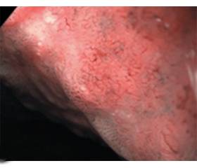
Актуальність. Стаття присвячена вивченню ксантом шлунка — доброякісних випадкових утворень з невстановленим етіопатогенезом, які пов’язані, за даними літератури, з низкою патологічних станів (атрофічний гастрит, кишкова метаплазія, дисплазія, рак шлунка, дисліпідемія тощо). Мета: вивчити частоту ксантом шлунка в пацієнтів з атрофічним гастритом й проаналізувати взаємозв’язок з ендоскопічними, у тому числі передраковими, змінами в шлунку. Матеріали та методи. Проведено езофагогастродуоденоскопію з використанням відеоендоскопічної системи EVIS EXERA III з гастроскопом Olympus 190 (Японія) 120 пацієнтам з атрофічним гастритом, які проходили обстеження й лікування в ДУ «Інститут гастроентерології НАМН України». Оцінювали наявність дуоденогастрального рефлюксу, поширеність і вираженість атрофії слизової оболонки (СО), зміни рельєфу СО (у тому числі наявність вузлувато-бугристого рельєфу), наявність гіперплазій, поліпів, геморагій, ерозій, виразок, ксантом у шлунку. При застосуванні функції вузькосмугової візуалізації (NBI) і ближнього фокуса оцінювали наявність і поширеність кишкової метаплазії, наявність дисплазії СО. Проводили порівняльну характеристику ендоскопічних даних залежно від наявності ксантом у шлунку, виконували статистичний аналіз отриманих даних. Результати. Встановлено неоднорідність ендоскопічних змін при атрофічному гастриті (поширення і вираженість атрофії, кишкової метаплазії, зміни рельєфу СО, наявність гіперплазій, поліпів, виразок тощо). Ксантоми шлунка виявлено більше ніж у третини хворих на атрофічний гастрит (36,7 % випадків) з переважною локалізацією в антральному відділі (90,9 %). Утворення, як правило, були поодинокими (95,5 %). У пацієнтів з атрофічним гастритом, асоційованим із ксантомами, значно частіше відзначались дифузні атрофічні зміни СО шлунка, були значно більшими частота й поширеність вузлувато-бугристого рельєфу СО і кишкової метаплазії, частота гіперплазій і дисплазій СО, що підтверджено морфологічно (p < 0,05). Поодинокі випадки виразок шлунка й раку шлунка діагностовано тільки в пацієнтів із ксантомами (p > 0,05). Висновки. Отримані результати свідчать про те, що ксантоматоз шлунка виступає в ролі важливого діагностичного маркера передракових змін СО шлунка (поширеної атрофії, кишкової метаплазії, дисплазії) і може бути важливим прогностичним маркером розвитку раку шлунка. Пацієнти з атрофічним гастритом, асоційованим із ксантомами, потребують ретельного обстеження із застосуванням сучасного ендоскопічного обладнання і динамічного спостереження із зосередженням особливої уваги на змінах рельєфу шлунка за типом вузлувато-бугристого.
Background The article deals with the study of gastric xanthomas, benign accidental formations with an unknown etiopathogenesis, which, according to the literature, are associated with a number of pathological conditions (atrophic gastritis, intestinal metaplasia, dysplasia, gastric cancer, dyslipidemia, etc.). The purpose was to study the prevalence of gastric xanthomas in patients with atrophic gastritis and to analyze their relationship with endoscopic changes in the stomach, including precancerous conditions. Materials and methods. Esophagogastroduodenoscopy was performed using the EVIS EXERA III video endoscopy system with the Olympus 190 gastroscope (Japan) in 120 patients with atrophic gastritis undergoing examination and treatment at the Institute of Gastroenterology of the National Academy of Sciences of Ukraine. The presence of duodenogastric reflux, the prevalence and severity of mucosal atrophy, changes in the relief of the gastric mucosa (including nodules and bumps), the presence of hyperplasia, polyps, hemorrhages, erosions, ulcers, xanthomas in the stomach were assessed. When using the narrow-band imaging and close focus, the presence and prevalence of intestinal metaplasia, the presence of gastric dysplasia were evaluated. Comparative and statistical analysis of endoscopic data was conducted depending on the presence of gastric xanthomas. Results. The heterogeneity of endoscopic changes in atrophic gastritis was revealed (prevalence and severity of atrophy, intestinal metaplasia, changes in the mucosal relief, presence of hyperplasia, polyps, ulcers, etc.). Gastric xanthomas were found in more than a third of patients with atrophic gastritis (36.7 % of cases), with predominant localization in the antral region (90.9 %). As a rule (95.5 %), they were single. In patients with atrophic gastritis associated with xanthomas, diffuse atrophic changes in gastric mucosa were detected significantly more often, as well as mucosal nodules and bumps, mucosal hyperplasia, intestinal metaplasia and the mucosal dysplasia, which was confirmed morphologically (p < 0.05). Rare cases of gastric ulcers and gastric cancer were diagnosed only in patients with xanthomas (p > 0.05). Conclusions. The obtained results indicate that gastric xanthoma is an important diagnostic marker of precancerous changes in the gastric mucosa: widespread atrophy, intestinal metaplasia, dysplasia, and can be used as an important prognostic marker for the development of gastric cancer. Patients with xanthoma-associated atrophic gastritis require a thorough examination using modern endoscopic equipment and dynamic observation, with a focus on changes in the gastric relief by the type of nodules and bumps.
ендоскопічне дослідження шлунка; ксантоми шлунка; атрофічний гастрит; кишкова метаплазія; дисплазія
upper endoscopy; gastric xanthomas; atrophic gastritis; intestinal metaplasia; dysplasia
Для ознакомления с полным содержанием статьи необходимо оформить подписку на журнал.
- Management of precancerous conditions and lesions in the stomach (MAPS): guideline from the European Society of Gastrointestinal Endoscopy (ESGE), European Helicobacter Study Group (EHSG), European Society of Pathology (ESP), and the Sociedade Portuguesa de Endoscopia Digestiva (SPED). Guideline / M. Dinis-Ribeiro et al. Endoscopy. 2012. Vol. 44. № 1. P. 74-94.
- Gastric Xanthomas and Fundic Gland Polyps as Endoscopic Risk Indicators of Gastric Cancer / K. Yamashita et al. Gut Liver. 2019. Vol. 13. № 4. P. 409-414.
- Степанов Ю.М., Сімонова О.В., Мосійчук Л.М., Петишко О.П. Сучасні ендоскопічні критерії стратифікації хворих на атрофічний гастрит при розвитку передракових змін шлунка. Патологія. 2018. Т. 15. № 3(44). С. 346-353.
- Chen Y., He X.-J., Zhou M.-J., Li Y.-M. Gastric xanthelasma and metabolic disorders: A large retrospective study among Chinese population. World J Gastroenterol. 2017. Vol. 23(43). P. 7756-7764.
- Gastric xanthalesmas: Report of five cases and review of the literature / O.S. Dizdar et al. J Exp Clin Med. 2015. Vol. 32. № 2. P. 91-95.
- Alzahrani M., Alqunaitir A., Alsohaibani M., Al-Rikabi A.C. Gastric xanthelasma associated with hyperplastic polyp and mucosal erosions: report of an unusual case and literature review. Oxf Med Case Reports. 2018. № 2018(8). omy051.
- Dhakal M., Dhakal O.P., Bhandari D., Gupta A. Gastric xanthelasma: an unusual endoscopic finding. BMJ case reports. Vol. 2013. P. bcr2013201017.
- Fukuda S., Akahoshi K., Fushimi F., Oya M. Gastric hyperplastic polyp with xanthoma observed by magnification narrow-band imaging endoscopy and endoscopic ultrasonography: report of a case. Fukuoka Acta Med. 2015. Vol. 106. № 4. P. 77-82.
- Moumin F.A., Mohamed A.A., Osman A.O., Cai J. Gastric Xanthoma Associated with Gastric Cancer Development: An Updated Review. Can J Gastroenterol Hepatol. 2020. № 2020. P. 3578927.
- Orth J. Lehrbuch specielle pathologische anatomie und histology, Berlin, 1887 (cited in Khachaturian T., Dinning J.P., Earnest D.L.). Gastric xantelasma in patient after partial gastrectomy. Am J Gastroenterol. 1998. 93. 1588-1589.
- Gencosmanoglu R., Sen-Oran E., Kurtkaya-Yapicier O., Tozun N. Xanthelasmas of the upper gastrointestinal tract. J Gastroenterol. 2004. Vol. 39. № 3. P. 215-219.
- Greenberg M.R., Shah Sh. An Unusual Case of a Gastric Xanthoma: A Case Report Cureus. 2022. Vol. 14. № 5. P. e25026.
- Ксантоми шлунка: випадкова знахідка чи важливий діагностичний маркер? / О.В. Сімонова та ін. Український журнал малоінвазивної та ендоскопічної хірургії. 2010. Т. 14. № 2. С. 45-46.
- Wu J.-H. Gastric Xanthomatosis: A Rare Presentation of a Common Disorder. Clin Gastroenterol Hepatol. 2016. Vol. 14. № 6. P. A18.
- Gastric Xanthoma Is Related to the Rapid Growth of Gastric Cancer / K. Miura et al. J Clin Med. 2021. № 10. P. 5704.
- Gastric xanthomas in the elderly / M. Naito et al. Nihon Ronen Igakkai Zasshi. 1991. Vol. 28. № 5. P. 683-687.
- Gastric xanthelasma may be a warning sign for the presence of early gastric cancer / A. Sekikawa et al. J Gastroenterol Hepatol. 2014. Vol. 29. № 5. P. 951-956.
- Gastric atrophy and xanthelasma are markers for predicting the development of early gastric cancer / A. Sekikawa et al. J Gastroenterol. 2016. Vol. 51. P. 35-42.
- A close relationship between Helicobacter pylori infection and gastric xanthoma / H. Isomoto et al. Scand J Gastroenterol. 1999. Vol. 34. № 4. P. 346-352.
- Hori S., Tsutsumi Y. Helicobacter pylori infection in gastric xanthomas: immunohistochemical analysis of 145 lesions. Pathology International. 1996. Vol. 46. № 8. P. 589-593.
- Is Gastric Xanthelasma an Alarming Endoscopic Marker for Advanced Atrophic Gastritis and Intestinal Metaplasia? / A.S. Köksal et al. Dig Dis Sci. 2016. Vol. 61. № 10. P. 2949-2955.
- Gastric xanthelasma may be a warning sign of intestinal metaplasia: A cross sectional study / D. Xiao et al. Oncol Rep. 2020. Vol. 44. № 3. P. 1275-1281.
- Type IIa early gastric cancer with proliferation of xanthoma cells / A. Muraoka et al. J Gastroenterol. 1998. Vol. 33. № 3. P. 326-329.
- Gastric Xanthoma Is a Predictive Marker for Early Gastric Cancer Detected after Helicobacter pylori Eradication / N. Shibukawa et al. Intern Med. 2019. Vol. 58. № 6. P. 779-784.