Інформація призначена тільки для фахівців сфери охорони здоров'я, осіб,
які мають вищу або середню спеціальну медичну освіту.

Підтвердіть, що Ви є фахівцем у сфері охорони здоров'я.

Журнал «Здоровье ребенка» Том 17, №5, 2022

Вернуться к номеру

Функціонально-біохімічна характеристика м’язової системи в дітей із цукровим діабетом 1-го типу

Авторы: G. Lezhenko (1), O. Pashkova (1), K. Samoylyk (1), A. Brutman (2)
(1) — Zaporizhzhia State Medical University, Zaporizhzhia, Ukraine
(2) — National University “Zaporizhzhia Polytechnic”, Zaporizhzhia, Ukraine

Рубрики: Педиатрия/Неонатология

Разделы: Справочник специалиста

Версия для печати


Резюме

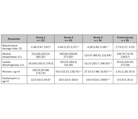
Мета: визначити можливі маркери ураження скелетних м’язів у дітей, хворих на цукровий діабет 1-го типу (ЦД1), та їх зв’язок з особливостями перебігу захворювання. Матеріали та методи. Групу спостереження становили 98 дітей із цукровим діабетом 1-го типу: 1-ша група включала 22 дитини без порушень з боку м’язової системи; 2-га — 42 пацієнти з динапенією; 3-тя — 34 дитини з діабетичною міопатією. Контрольна група — 30 умовно здорових дітей. Усім пацієнтам було проведено дослідження статичної витривалості скелетних м’язів, визначення рівня креатинкінази, аспартат­амінотрансферази, лужної фосфатази, лактатдегідрогенази, періостину та кардіотрофіну-1 у сироватці крові. Результати. Проведене дослідження показало, що в дітей, хворих на ЦД1, незалежно від структурно-функціонального стану м’язової системи, спостерігаються ознаки пошкодження скелетних м’язів, що були максимально вираженими при діабетичній міопатії та прогресували при погіршенні глікемічного конт­ролю. Указані порушення відбувалися на тлі змін активності лужної фосфатази, найбільші показники якої спостерігалися в 1-й групі, у той же час у пацієнтів 3-ї групи її вміст у сироватці крові відповідав значенням контрольної групи. Одночасно при розвитку діабетичної міопатії в дітей, хворих на ЦД1, активність лактатдегідрогенази зростала в 1,2 раза (p < 0,01) та кардіотрофіну-1 — у 300 разів (p < 0,01) порівняно з аналогічними показниками контрольної групи. Уміст періостину був підвищеним у всіх групах пацієнтів, хворих на ЦД1. Його значення були максимальними в 1-й групі пацієнтів, перевищуючи показники контрольної групи в 103 рази (p < 0,01). При погіршенні стану скелетної мускулатури рівень періостину в сироватці крові поступово знижувався, але при динапенії залишався вищим за його значення в групі контролю в 35,5 раза (p < 0,05), а при діабетичній міопатії — у 19,2 раза (p < 0,05). Висновки. Перебіг ЦД1 у дітей супроводжується ураженням скелетних м’язів, першою клінічною ознакою якого є зниження статичної витривалості скелетних м’язів, що відбувається на тлі погіршення перебігу захворювання. Біохімічними маркерами ураження скелетних м’язів у дітей, хворих на ЦД1, є лужна фосфатаза, лактатдегідрогеназа, періостін та кардіотрофін-1. Загальною рисою змін означених показників є їх зростання, однак кожному клінічному стану скелетної мускулатури відповідає своя конфігурація змін вказаних маркерів.

Background. The purpose of the study was to determine possible markers of skeletal muscle damage in children with type 1 diabetes mellitus (T1DM) and their relationship with the features of disease course. Materials and methods. The observation group consisted of 98 children with type 1 diabetes mellitus: the first group included 22 people without disorders of the muscular system; the second — 42 patients with dynapenia; the third — 34 children with diabetic myopathy. Control group — 30 relatively healthy children. Assessment of the static endurance of skeletal muscles, determination of the level of creatine kinase, aspartate aminotransferase, alkaline phosphatase, lactate dehydrogenase, periostin and cardiotrophin-1 in blood serum were performed in all patients. Results. The conducted studies demonstrate that children with diabetes, regardless of the structural and functional state of their muscular system, have signs of skeletal muscle damage, which were most expressed in diabetic myopathy and progressed with maximal deterioration of glycemic control. It was found that the highest content of alkaline phosphatase was characteristic of children from group 1, while in patients with diabetic myopathy its serum content was not statistically different from that of controls. These disorders occurred against the background of changes in alkaline phosphatase activity, the level of which was highest in children from group 1, while in patients with diabetic myopathy, its serum content was not statistically different from that of controls. At the same time, during the course of diabetic myopathy in children with T1DM, there was an increase in lactate dehydrogenase activity by 1.2 times (p < 0.01) and cardiotrophin-1 by 300 times (p < 0.01) compared to the corresponding indicator of the control group. Serum periostin level was increased in all patients with T1DM. Its maximum values were determined in group 1, whose periostin concentration exceeded control indicators by 103 times (p < 0.01). With deterioration of skeletal muscle state, there was a gradual decrease in periostin serum level, but in patients with dynapenia, it was 35.5 times higher than in the control group (p < 0.05) and 19.2 times higher in those with diabetic myopathy (p < 0.05). Conclusions. The course of type 1 diabetes in children is accompanied by skeletal muscle damage, the first clinical sign of which is a decrease in the static muscle endurance against the background of worsening disease course. Alkaline phosphatase, lactate dehydrogenase, periostin, and cardiotrophin-1 are biochemical markers of skeletal muscle damage in children with type 1 diabetes. A common feature of the changes in the specified indicators is their increase; however, each clinical condition of the skeletal muscles corresponds to its own configuration of changes in the abovementioned markers.


Ключевые слова

діти; цукровий діабет 1-го типу; діабетична міопатія; маркери ураження скелетних м’язів

children; type 1 diabetes; diabetic myopathy; markers of skeletal muscle damage

Introduction

Skeletal muscles make up approximately 40 % of our body weight and are one of largest organs in terms of mass and protein content. However, the mass of skeletal muscles is very dynamic and depends on both physiological and pathological conditions. Regulation of muscle mass, determining their growth or atrophy, depends on the balance between anabolic and catabolic stimuli [1]. Loss of skeletal muscle mass and decline in muscle function are hallmarks of aging. This process reaches its maximum on the seventh and eighth decades of life. However, in people with type 1 diabetes (T1DM), diabetes-related decrease in muscle mass begins at a much younger age [2]. In addition to impaired muscle growth and function, in T1DM there is a deterioration of their recovery after damage, which may be associated with the loss of muscle stem cells [3]. This condition in diabetic patients is called diabetic myopathy, it can be observed in 27–88 % of cases [4, 5]. This serious, often overlooked complication is believed to contribute to the progression of chronic diabetic complications due to the key role of skeletal muscle in glucose homeostasis [6, 7]. Despite the fact that patients with T1DM strive to maintain normal glycemic control, less than a third of them consistently achieve target blood glucose levels [8]. Hyperglycemia and the resulting advanced glycation end products are leading factors of muscle dysfunction in diabetes and may significantly contribute to the development of diabetic myopathy in T1DM [9]. Chronic low-grade inflammation inherent in T1DM patients impairs skeletal muscle regeneration [10]. The findings of the 2020 meta-analysis conclusively demonstrate a significant association between higher levels of circulating inflammatory markers and lower skeletal muscle strength and muscle mass [11]. Tumor necrosis factor, interleukin 6, interleukin 1, and interferon γ are among the most studied proinflammatory cytokines contributing to the development of muscle atrophy [12]. Important mechanisms in the pathogenesis of diabetic myopathy are increased oxidative stress and impaired antioxidant protection [13]. Recent studies have shown that alteration within the mitochondrial oxidative capacity of young adults with type 1 diabetes can be a possible mechanism of muscle fatigue [14]. The roles of insulin [15], insulin-like growth factor 1 and 2 [16, 17], basic fibroblast growth factor [18], transforming growth factor β [19] and other myokines in skeletal myogenesis were also confirmed. However, despite the significant interest of scientists in the problems of skeletal muscle changes in diabetes and their role in the course of the disease, the issue of early manifestations of diabetic myopathy and markers of skeletal muscle damage in children with T1DM remains poorly studied.
The purpose of the study: to determine possible markers of skeletal muscle damage in children with type 1 diabetes and their relationship with the features of disease course.

Materials and methods

The study was conducted at the endocrinology department of the Municipal Institution “Zaporizhzhia Regional Clinical Children’s Hospital” of the Zaporizhzhia Regional Council and involved 98 children with type 1 diabetes aged 11 to 17 years. Depending on the condition of the skeletal muscles, patients were divided into 3 groups. The first group included 22 children without muscular disorders. The second group consisted of 42 children whose muscular system condition corresponded to dynapenia. The third group included 34 patients diagnosed with diabetic myopathy. The control group consisted of 30 conditionally healthy children. All groups were representative of age, sex, body mass index, and duration of diabetes. Exclusion criteria: the absence of consent to participate in the study; obesity or overweight; the presence of acute inflammatory processes or congenital malformations in the stage of decompensation; professional engagement in sports.
Diabetic myopathy was diagnosed when the hand strength index was less than 49.6 % in boys, less than 43 % in girls, and the skeletal muscle index was less than 75.3 %, regardless of gender. Dynapenia was detected with a reduction of only the wrist strength index when determining the skeletal muscle index of 75.3 % and above [4].
All children underwent the measuring of body weight and height with the further evaluation of body mass index. The muscular mass in patients under 15 years of age was estimated according to A.M. Peters equation [20]. P. Boer equation was applied for children above 15 years of age taking into account gender [21]. To evaluate condition of muscular system, the skeletal muscle index was assessed [22]. Body fat percentage [23] and body fat mass were also determined [24].
Skeletal muscle strength was assessed using a hand spring dynamometer DK-50. To level out the age of a child when estimating the muscle strength, one has applied the wrist strength index (WSI). WSI was evaluated by the following equation:
WSI = (wrist strength (kg) / body mass (kg)) × 100 %.
To define functional abilities of muscular system, static endurance of skeletal muscles was estimated while fixing the maximal period of sustaining a given position in seconds. A series of tests was performed to determine static endurance of the following muscles: 1) neck flexors — the child was offered to keep his head raised as long as possible in a position lying on his back to a height limited by the distance of the chest from the couch; 2) back extensors: the starting position — lying on the stomach, the legs are fixed, the chest is kept raised above the couch as long as possible, with hands behind the head; 3) abdominal muscles: starting position — lying on the back, arms along the body, raise the legs to an angle of 45° and hold for the maximum time; 4) gluteus medius — initial standing position, maximum adduction of the hip to the side, prevention of its rotation. We also evaluated the total static endurance of the muscles as the sum of the time of static endurance of all muscle groups studied [4].
All children underwent a biochemical blood analysis to determine the level of fasting blood glucose, creatine kinase, aspartate aminotransferase, alkaline phosphatase, and lactate dehydrogenase (LDH). Muscle tissue damage index (MTDI) was determined as the creatine kinase to aspartate aminotransferase ratio.
The content of serum periostin was determined by the method of enzyme immunoassay using the Human Periostin/OSF-2 ELISA Kit. Plasma level of cardiotrophin-1 was measured with enzyme immunoassay using the RayBio® Human CT-1 ELISA Kit.
All the results were analyzed using the set of statistic programs Statistica 13.0 (StatSoft Inc., No. JPZ8041382130ARCN10-J), with the Shapiro-Wilk asymmetry test of normality. To compare characteristics, the median (Me) and quartiles of Me (Q25; Q75) were used. The reliability of the differences in the obtained results for different groups was determined by the Student’s test. Correlations were evaluated by the Pearson coefficient. Differences were considered to be significant at p < 0.05.
When planning this work, we obtained a permission from the regional bioethical commission of Zaporizhzhia State Medical University. All procedures performed in studies involving children conformed to the ethical standards of the institutional and national research committees and the 1964 Declaration of Helsinki and its later amendments or comparable ethical standards. Informed consent was obtained from each of the study participants and their official guardians.

Results

In all groups of children with diabetes, including patients of group 1, whose muscle mass and skeletal muscle strength remained preserved, the static endurance tests of the studied skeletal muscle revealed a deterioration in the ability to maintain given posture as much as possible, which led to a statistically significant decrease in total static endurance compared to the similar indicator of the control group (Table 1).
The results of total static muscle endurance were worst in children, who didn’t achieve optimal glycemic control (the correlation coefficient between the total static muscle endurance and glycated hemoglobin was r = –0.47, p < 0.05). This occurred against the background of skeletal muscle damage, confirmed by an increase in MTDI, which was 1.2 times higher among patients in group 1 than in controls (p < 0.05). The highest MTDI was observed in the group of children with dynapenia and diabetic myopathy. Its value exceeded the indicators not only of the control group, but also of group 1 (p < 0.05) (Table 2).
Further, we investigated the content of alkaline phosphatase, the activity of which, according to literature data, changes in skeletal muscle pathology [25]. The highest levels of alkaline phosphatase were found in children from group 1, while in patients with diabetic myopathy, its serum content was not statistically different from the control group (Table 2). Simultaneously with a decrease in the content of alkaline phosphatase against the background of a damage to the structural and functional muscles, there was an increase in LDH activity, which plays an important role in ensuring the normal physiology of skeletal muscles [26], with its maximum value in group 3 (Table 2).
Given that periostin is necessary for maintaining muscle mass during muscle regeneration and plays protective role in muscular dystrophy [27], we investigated its serum level in children with diabetes, depending on structural and functional state of skeletal muscles (Table 2). The obtained results showed elevated periostin content in all groups of patients with diabetes, compared to indicators in controls. Its maximum values were determined in group 1 patients, whose periostin concentration exceeded that of controls by 103 times (p < 0.01). With structural and functional changes in the skeletal muscles, there was a gradual decrease in periostin serum level. Thus, in patients with dynapenia, its content was 35.5 times higher than in the control group (p < 0.05) and 19.2 times higher in people with diabetic myopathy (p < 0.05). An inverse relationship between serum glucose content and periostin level was established (r = –0.48, p < 0.05)
The highest level of cardiotrophin-1, which according to the literature is involved in the development of skeletal muscle atrophy [28], was found in patients with diabetic myopathy. It exceeded that of the control group by a median of 300 times (p < 0.01). Among the patients in groups 1 and 2, the content of cardiotrophin-1 was also statistically higher than in controls (p < 0.01), but compared to group 3, it was 11.5 and 8 times lower, respectively (p < 0.05). The highest values of cardiotrophin-1 were determined in children with high blood glucose levels (r = +0.48, p < 0.05).

Discussion

The conducted studies demonstrate that children with diabetes, regardless of the structural and functional state of the muscular system, have signs of skeletal muscle damage, as evidenced by an increase in MTDI. In physiological conditions, the skeletal muscle is a stable structure. However, in case of its damage, activation of satellite cells — resident somatic stem muscle cells — occurs to restore structural integrity. In chronic muscle damage observed in diabetes, the cycles of muscle damage and regeneration are constantly repeated. This leads to changes in the architectonics of skeletal muscle tissue, causing fibrosis, fatty infiltration, myofibrillar atrophy [29], which is a manifestation of diabetic myopathy [4].
These violations occurred against the background of changes in the activity of alkaline phosphatase, one of whose biological functions is active transport of metabolites through biological membranes [30]. Therefore, the established decrease in serum content of this enzyme with the progression of structural and functional changes in skeletal muscles of children with diabetes seems logical. It is possible that an increased activity of alkaline phosphatase in patients with preserved mass and strength of skeletal muscles may be due to the development of nonspecific low-grade inflammation and oxidative stress against the background of hyperglycemia [31], which acts as a factor of skeletal muscle fibrosis [32] and a trigger for muscle loss [25]. As chronic oxidative stress progresses, alkaline phosphatase activity decreases. Alkaline phosphatase deficiency causes loss of skeletal muscle mass, muscle strength, and increased fatigue due to the direct effect on muscle and neuronal progenitor cells, their development, and function [33–35]. It is known that alkaline phosphatase is an endothelial marker of the microcirculatory system, and its decrease in diabetic patients, to a certain extent, may indicate a microvascular alterations [36]. This assumption is consistent with our previous studies revealing a latent disorder of peripheral circulation during the development of diabetic myopathy [37]. This and, as a result, hypoxemia of tissues, including muscles, can be the cause of deterioration of functional capacity of muscles [5].
Significant biochemical changes in children with diabetes were also manifested by an increase in LDH level, as the structural and functional changes in the skeletal muscles progressed. Selective increase in LDH level, which reflects the activity of the glycolytic pathway of metabolism, may be a response to chronic oxidative stress in children with diabetes [4] since LDH is known to be involved in antioxidant protection [26, 38]. On the other hand, LDH accumulation leads to the destruction of cellular structures and degenerative changes in myofibrils [30].
We also observed high levels of periostin in diabetic children, with the highest values in patients with preserved muscle mass and strength. Our data, as other studies, have demonstrated that periostin expression is very low in intact muscle [27]. However, when skeletal muscles are damaged, secretion of periostin increases [39]. It participates in tissue regeneration and remodeling of skeletal muscles [40]. It is believed that high glucose and inflammatory cytokine concentrations may promote the regulation of periostin biosynthesis in target tissues [41]. At the first stages, muscle regeneration under the influence of periostin occurs without fibrosis. With long-term chronic muscle damage induced by high glucose level and repeated muscle regeneration, activation of periostin synthesis leads to irreversible fibrotic processes in skeletal muscles. As a result, functional capabilities are impaired and the process of myofibril regeneration is inhibited, which subsequently leads to a decrease in periostin synthesis [29]. Experimental studies found that in the absence of periostin in animals, there was a decrease in muscle mass due to the loss of muscle fibers during repeated regeneration. The authors related the loss of muscle fibers not to an impaired function of muscle stem cells, but to a deterioration in the supply of nutrients from blood vessels due to a decrease in their number. This indicates the role of periostin in the regulation of angiogenesis during muscle regeneration [27].
Cardiotrophin-1, a member of the interleukin-6 cytokine family, is also a powerful inhibitor of skeletal muscle differentiation and regeneration. The main sites of cardiotrophin-1 expression during embryonic development are the heart and skeletal muscles [42]. Experimental works established that activation and proliferation of satellite cells occurs when myofibrils are damaged under the action of cardiotrophin-1. As a result, many daughter myoblasts are formed at the site of injury to renew or replace lost myofibrils [43]. At the same time, cardiotrophin-1 maintains an undifferentiated state in muscle progenitor cells and delays the regeneration of damaged muscles in vivo [42, 43]. Thus, an elevated level of cardiotrophin-1 in children with diabetes can be a risk factor for the development of diabetic myopathy. This assumption was confirmed by the results of our study, which indicated that the progression of structural and functional changes in the skeletal muscles of children with diabetes was associated with increased content of cardiotrophin-1, with its maximum values in the group of patients with diabetic myopathy. Also, plasma cardiotrophin-1 concentration is associated with indicators of vascular diseases. The work of Gamella-Pozuelo L. et al. (2015) has found a negative correlation between cardiotrophin-1 and the ankle-brachial index in patients with diabetes [44]. This is consistent with the results of our previous studies, which proved the role of peripheral blood circulation disorders in the development of diabetic myopathy [4, 37]. At the same time, a direct correlation between cardiotrophin-1 and glucose indicates that activation of cardiotrophin-1 synthesis may be associated with hyperglycemia. Similar conclusions were reached by other researchers, in whose works it was proved that cardiotrophin-1 has hypoglycemic properties by stimulating glucose absorption in myofibrils [45]. Moreover, the study of Moreno-Aliaga et al. (2011) has proved insulin-independent effect of cardiotrophin-1 on glucose metabolism [46].

Conclusions

1. The course of type 1 diabetes in children is accompanied by skeletal muscle damage, the first clinical sign of which is a decrease in the static muscle endurance against the background of worsening disease course and, respectively, an increase in the glycated hemoglobin level.
2. Alkaline phosphatase, lactate dehydrogenase, periostin, and cardiotrophin-1 are biochemical markers of skeletal muscle damage in children with type 1 diabetes. A common feature of changes in the specified indicators is their increase; however, each clinical condition of the skeletal muscles corresponds to its own configuration of changes in the abovementioned markers.
 
Received 13.08.2022
Revised 21.08.2022
Accepted 27.08.2022

Список литературы

  1. Daou H.N. Exercise as an anti-inflammatory therapy for cancer cachexia: a focus on interleukin-6 regulation. American Journal of Physiology-Regulatory, Integrative and Comparative Physiology. 2020. 318(2). R296-R310. doi: 10.1152/ajpregu.00147.2019.
  2. Monaco C.M., Gingrich M.A., Hawke T.J. Considering type 1 diabetes as a form of accelerated muscle aging. Exercise and Sport Sciences Reviews. 2019. 47(2). 98-107. doi: 10.1249/JES.0000000000000184.
  3. Monaco C.M., Perry C.G., Hawke T.J. Diabetic myopathy: current molecular understanding of this novel neuromuscular disorder. Current Opinion in Neurology. 2017. 30(5). 545-552. doi: 10.1097/WCO.0000000000000479.
  4. Chudova N.I. Early diagnosis, prediction and objectives of approaches to the prevention of muscular system disorders in children suffering from diabetes mellitu. PhD Thesis. Zaporizhzhia State Medical University of the Ministry of Health of Ukraine. Zaporizhzhia, 2022. Available from: https://zsmu.edu.ua/upload/updisert/dfpediatr/12022022_dis_chudova.pdf.
  5. Zhurakivska O.Y., Koshkin O.Y., Tkachuk Y.L., Knyazevych-Chorna T.V., Rudyak O.M. Age characteristics of morphogenesis of diabetic myopathies. Problems of Endocrine Pathology. 2020. 74(4). 115-123. doi: 10.21856/j-PEP.2020.4.15.
  6. Coleman S.K., Rebalka I.A., D’Souza D.M., Hawke T.J. Skeletal muscle as a therapeutic target for delaying type 1 diabetic complications. World Journal of Diabetes. 2015. 6(17). 1323. doi: 10.4239/wjd.v6.i17.1323.
  7. Dimitriadis G.D., Maratou E., Kountouri A., Board M., Lambadiari V. Regulation of postabsorptive and postprandial glucose metabolism by insulin-dependent and insulin-independent mechanisms: an integrative approach. Nutrients. 2021. 13(1). 159. doi: 10.3390/nu13010159.
  8. Type 1 Diabetes Statistics. Available from: https://beyondtype1.org/type-1-diabetes-statistics/.
  9. Travis C., Srivastava P.S., Hawke T.J., Kalaitzoglou E. Diabetic bone disease and diabetic myopathy: manifestations of the impaired muscle-bone unit in type 1 diabetes. Journal of Diabetes Research. 2022. 2022. doi: 10.1155/2022/2650342.
  10. Perandini L.A., Chimin P., Lutkemeyer D. da S., Câmara N.O.S. Chronic inflammation in skeletal muscle impairs satellite cells function during regeneration: can physical exercise restore the satellite cell niche? The FEBS Journal. 2018. 285(11). 1973-1984. doi: 10.1111/febs.14417.
  11. Tuttle C.S., Thang L.A., Maier A.B. Markers of inflammation and their association with muscle strength and mass: A systematic review and meta-analysis. Ageing Research Reviews. 2020. 64. 101185. doi: 10.1016/j.arr.2020.101185.
  12. Haberecht-Müller S., Krüger E., Fielitz J. Out of control: the role of the ubiquitin proteasome system in skeletal muscle during inflammation. Biomolecules. 2021. 11(9). 1327. doi: 10.3390/biom11091327.
  13. Jurisic-Erzen D., Starcevic-Klasan G., Ivanac D., Peharec S., Girotto D., Jerkovic R. The effects of alpha-lipoic acid on diabetic myopathy. Journal of Endocrinological Investigation. 2018. 41(2). 203-209. doi: 10.1007/s40618-017-0720-0.
  14. Monaco C.M., Hughes M.C., Ramos S.V., Varah N.E., Lamberz C., Rahman F. A., Perry C.G. Altered mitochondrial bioenergetics and ultrastructure in the skeletal muscle of young adults with type 1 diabetes. Diabetologia. 2018. 61(6). 1411-1423. doi: 10.1007/s00125-018-4602-6.
  15. Turner M.C., Player D.J., Martin N.R., Akam E.C., Lewis M.P. The effect of chronic high insulin exposure upon metabolic and myogenic markers in C2C12 skeletal muscle cells and myotubes. Journal of Cellular Biochemistry. 2018. 119(7). 5686-5695. doi: 10.1002/jcb.26748.s.
  16. Ahmad S.S., Ahmad K., Lee E.J., Lee Y.H., Choi I. Implications of insulin-like growth factor-1 in skeletal muscle and various diseases. Cells. 2020. 9(8). 1773. doi: 10.3390/cells9081773.
  17. Torrente Y., Bella P., Tripodi L., Villa C., Farini A. Role of insulin-like growth factor receptor 2 across muscle homeostasis: Implications for treating muscular dystrophy. Cells. 2020. 9(2). 441. doi: 10.3390/cells9020441.
  18. Jin B., Zhang L., Wang X., Jin D. Research on orientation of basic fibroblast growth factor with magnetic nanoparticles (MNPs) on regeneration and recovery of rats’ dampened skeletal muscle and expressed level of matrix metalloproteinase. Journal of Biomedical Nanotechnology. 2022. 18(2). 557-564. doi: 10.1166/jbn.2022.3260.
  19. Ismaeel A., Kim J.S., Kirk J.S., Smith R.S., Bohannon W.T., Koutakis P. Role of transforming growth factor-β in skeletal muscle fibrosis: a review. International Journal of Molecular Sciences. 2019. 20(10). 2446. doi: 10.3390/ijms20102446.
  20. Peters A.M., Snelling H.L.R., Glass D.M., Bird N.J. Estimation of lean body mass in children. Survey of Anesthesiology. 2012. 56(1). 26-27. doi: 10.1097/01.SA.0000410700.55371.0f.
  21. Boer P. Estimated lean body mass as an index for normalization of body fluid volumes in humans. American Journal of Physiology-Renal Physiology. 1984. 247(4). F632-F636. doi: 10.1152/ajprenal.1984.247.4.F632.
  22. Janssen I., Heymsfield S.B., Ross R. Low relative skeletal muscle mass (sarcopenia) in older persons is associated with functional impairment and physical disability. Journal of the American Geriatrics Society. 2002. 50(5). 889-896. doi: 10.1046/j.1532-5415.2002.50216.x.
  23. Deurenberg P., Weststrate J.A., Seidell J.C. Body mass index as a measure of body fatness: age -and sex-specific prediction formulas. British Journal of Nutrition. 1991. 65(2). 105-114. doi: 10.1079/bjn19910073.
  24. Akay A.F., Gedik A., Tutus A., Şahin H., Bircan M.K. Body mass index, body fat percentage, and the effect of body fat mass on SWL success. International Urology and Nephrology. 2007. 39(3). 727-730. doi: 10.1007/s11255-006-9133-2.
  25. Lee J.H., Cho A.R., Lee Y.J. Relationship between serum alkaline phosphatase and low muscle mass index among Korean adults: a nationwide population-based study. Biomolecules. 2021. 11(6). 842. doi: 10.3390/biom11060842.
  26. Young A., Oldford C., Mailloux R.J. Lactate dehydrogenase supports lactate oxidation in mitochondria isolated from different mouse tissues. Redox Biology. 2020. 28. 101339. doi: 10.1016/j.redox.2019.101339.
  27. Ito N., Miyagoe-Suzuki Y., Takeda S.I., Kudo A. Periostin is required for the maintenance of muscle fibers during muscle regeneration. International Journal of Molecular Sciences. 2021. 22(7). 3627. doi: 10.3390/ijms22073627.
  28. Tanaka T., Narazaki M., Kishimoto T. IL-6 in inflammation, immunity, and disease. Cold Spring Harbor Perspectives in Biology. 2014. 6(10). a016295. doi: 10.1101/cshperspect.a016295.
  29. Özdemir C., Akpulat U., Sharafi P., Yıldız Y., Onbaşılar İ., Kocaefe Ç. Periostin is temporally expressed as an extracellular matrix component in skeletal muscle regeneration and differentiation. Gene. 2014. 553. 130-139. doi: 10.1016/j.gene.2014.10.014.
  30. Raskaliei T.Y., Raskaliei V.B., Shobat L.B., Gavriliuk-Skiba G.O. Histochemical study of skeletal muscles in experimental spinal cord blunt injury. Morphologia. 2016. 10(3). 243-247. doi: 10.26641/1997-9665.2016.3.243-247.
  31. Pan W., Miao L., Lin Y., Huang X., Ge X., Moosa S. L., Pan L. Regulation mechanism of oxidative stress induced by high glucose through PI3K/Akt/Nrf2 pathway in juvenile blunt snout bream (Megalobrama amblycephala). Fish & Shellfish Immunology. 2017. 70. 66-75. doi: 10.1016/j.fsi.2017.09.005.
  32. Arnò B., Galli F., Roostalu U., Aldeiri B.M., Miyake T., Albertini A., Cossu G. TNAP limits TGF-β-dependent cardiac and skeletal muscle fibrosis by inactivating the SMAD2/3 transcription factors. Journal of Cell Science. 2019. 132(15). jcs234948. doi: 10.1242/jcs.234948.
  33. Zhang Z., Nam H.K., Crouch S., Hatch N.E. Tissue nonspecific alkaline phosphatase function in bone and muscle progenitor cells: Control of mitochondrial respiration and ATP production. International Journal of Molecular Sciences. 2021. 22(3). 1140. doi: 10.3390/ijms22031140.
  34. Ohlebusch B., Borst A., Frankenbach T., Klopocki E., Jakob F., Liedtke D., Graser S. Investigation of alpl expression and Tnap-activity in zebrafish implies conserved functions during skeletal and neuronal development. Scientific Reports. 2020. 10(1). 1-16. doi: 10.1038/s41598-020-70152-5.
  35. Liedtke D., Hofmann C., Jakob F., Klopocki E., Graser S. Tissue-nonspecific alkaline phosphatase — a gatekeeper of physiological conditions in health and a modulator of biological environments in disease. Biomolecules. 2020. 10(12). 1648. doi: 10.3390/biom10121648.
  36. Pan Y., Dong Y., Hou W., Ji Z., Zhi K., Yin Z., Chen Y. Effects of PEMF on microcirculation and angiogenesis in a model of acute hindlimb ischemia in diabetic rats. Bioelectromagnetics. 2013. 34(3). 180-188. doi: 10.1002/bem.21755.
  37. Pashkova O., Chudova N. The role of perifheral circulation disorders in the development of diabetic myopathy in children with diabetes mellitus. Actual Problems of Modern Medicine. 2021. (8). 69-77. doi: 10.26565/2617-409X-2021-8-07.
  38. Lemire J., Auger C., Mailloux R., Appanna V.D. Mitochondrial lactate metabolism is involved in antioxidative defense in human astrocytoma cells. Journal of Neuroscience Research. 2014. 92(4). 464-475. doi: 10.1002/jnr.23338.
  39. Field S., Uyttenhove C., Stroobant V., Cheou P., Donckers D., Coutelier J.P., Jat P.S. Novel highly specific anti-periostin antibodies uncover the functional importance of the fascilin 1-1 domain and highlight preferential expression of periostin in aggressive breast cancer. International Journal of Cancer. 2016. 138(8). 1959-1970. doi: 10.1002/ijc.29946.
  40. Szyszka M., Skrzypczyk P., Stelmaszczyk-Emmel A., Pańczyk-Tomaszewska M. Serum Periostin as a potential biomarker in pediatric patients with primary hypertension. Journal of Clinical Medicine. 2021. 10(10). 2138. doi: 10.3390/jcm10102138.
  41. Luo Y., Qu H., Wang H., Wei H., Wu J., Duan Y., Deng H. Plasma periostin levels are increased in Chinese subjects with obesity and type 2 diabetes and are positively correlated with glucose and lipid parameters. Mediators of Inflammation. 2016. 2016. 6423637. doi: 10.1155/2016/6423637.
  42. Miyake T., Alli N.S., Aziz A., Knudson J., Fernando P., Megeney L.A., McDermott J.C. Cardiotrophin-1 maintains the undifferentiated state in skeletal myoblasts. Journal of Biological Chemistry. 2009. 284(29). 19679-19693. doi: 10.1074/jbc.M109.017319.
  43. Alli N.S. Role and regulation of FRA-2 during skeletal muscle development. Doctoral Dissertation, York University. Toronto, 2014.
  44. Gamella-Pozuelo L., Fuentes-Calvo I., Gomez-Marcos M.A., Recio-Rodriguez J.I., Agudo-Conde C., Fernández-Martín J.L., Martínez-Salgado C. Plasma cardiotrophin-1 as a marker of hypertension and diabetes-induced target organ damage and cardiovascular risk. Medicine. 2015. 94(30). doi: 10.1097/MD.0000000000001218.
  45. Escoté X., Gómez-Zorita S., López-Yoldi M., Milton-Laskibar I., Fernández-Quintela A., Martínez J.A., Portillo M.P. Role of omentin, vaspin, cardiotrophin-1, TWEAK and NOV/CCN3 in obesity and diabetes development. International Journal of Molecular Sciences. 2017. 18(8). 1770. doi: 10.3390/ijms18081770.
  46. Moreno-Aliaga M.J., Pérez-Echarri N., Marcos-Gómez B., Larequi E., Gil-Bea F.J., Viollet B., Bustos M. Cardiotrophin-1 is a key regulator of glucose and lipid metabolism. Cell Metabolism. 2011. 14(2). 242-253. doi: 10.1016/j.cmet.2011.05.013.

Вернуться к номеру